The University of Illinois Chicago team has introduced a breakthrough cell imaging technique called FINICI (Fluctuation Increase Negated by Intra-Chain Interaction) that addresses a persistent challenge in biosensor technology. Traditional negative biosensors have often been unusable because, as the sensors lose signal when activity is detected, regions of high enzyme activity can look identical to regions with no activity. This limitation stems from the fundamental design flaw where negative biosensors cannot distinguish between areas where enzymes are actively working and those where they are completely absent. FINICI resolves this issue by inverting the optical output of these sensors, exposing previously hidden enzyme activity with remarkable precision.
This innovation relies on modifying the electronic state of a chromophore—a fluorescent protein—through electrostatic interactions. The adjustment limits the chromophore’s movement, reducing fluorescence and blinking in inactive zones while maintaining a clear signal where enzymes are active. Gary Mo and Kriti Srivastava, who led the development, explained that this interaction “restricts the chromophore, disallows electron delocalization, and reduces the number of transitions possible,” which in turn suppresses fluorescence and blinking in inactive regions. As a result, FINICI can visualize cellular structures smaller than what conventional light microscopy’s diffraction limit allows, enabling researchers to observe activity in compartments that were previously invisible.
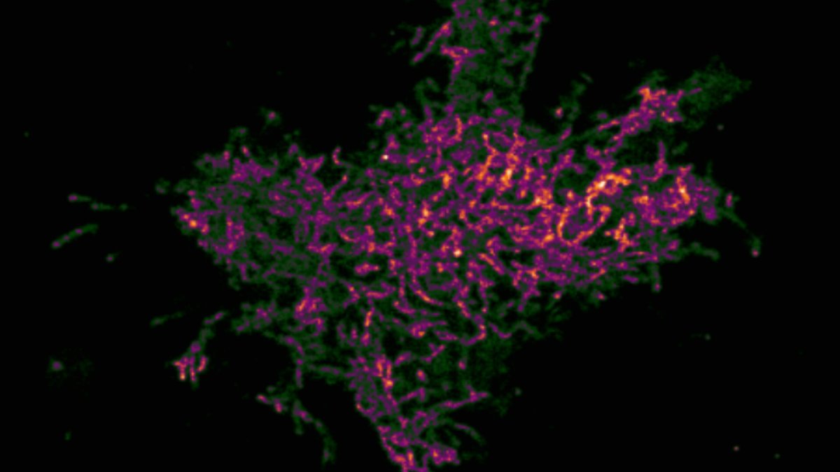
Gary Mo and Kriti Srivastava led the development and testing of FINICI across three enzymes: Src kinase, Syk kinase, and cGMP. Their experiments revealed that Src kinase, associated with cancer progression and cell movement, exhibited concentrated bursts of activity in specific membrane regions, including cholesterol-rich lipid rafts. Some of these active sites appeared briefly before disappearing, while others remained stable—details that conventional whole-cell measurements would miss entirely.
The researchers noted that these observations suggest membrane lipids may play a more dynamic role in enzyme anchoring than previously recognized, as Src kinase activity fluctuated even within the same lipid domain. Additionally, the team found that cGMP formed small clusters that were quickly overwhelmed as the signal spread, indicating that cells can dynamically regulate enzyme activity within nanoscale domains rather than relying solely on fixed localization.
Read Also: Novo Nordisk’s ziltivekimab fails Phase 3 trial
Syk kinase, which plays a key role in immune signaling, showed the highest activity near internal scaffolding structures within immune cells. Unlike Src kinase, Syk’s activity was concentrated away from the membrane, where adaptor proteins likely anchor it for activation. The team also found that cGMP formed small clusters that were quickly overwhelmed as the signal spread.
Mo and Srivastava said in an email: “From our perspective, the findings together showcased new ways that cells actively use compartmentalization. We knew that enzymes are anchored by adaptor proteins, and that appears true for Syk, only away from the membrane where it’s activated. But the case with Src hints that membrane lipids can also control anchoring. And cGMP showed that forming a nanodomain is not the end, we can still tune its usefulness via a dynamic background.”
FINICI outshines FRET in low-signal detection
When compared to Förster resonance energy transfer (FRET), the established benchmark in biosensing, FINICI demonstrated superior performance. In cells with low receptor expression, FINICI detected enzyme activity where FRET failed to register any signal. The researchers validated this sensitivity by directly comparing FINICI’s performance against FRET, confirming that FINICI consistently outperformed the gold standard in low-signal conditions. To ensure accuracy, the team employed biological controls, cells lacking the target enzyme, to confirm that FINICI’s readings were specific and not the result of false positives. This rigorous validation shows the technique’s reliability in distinguishing true enzymatic activity from background noise.
The potential impact of this method extends far beyond basic research. In drug development, FINICI could clarify whether enzymatic activity follows dispersed or localized patterns, whether it aligns with known cellular structures, and how individual cells respond differently. Such precision could enhance screening protocols, much like transcriptomic analysis deepens understanding of gene function. Mo and Srivastava highlighted that FINICI provides “previously hidden dimensions” in drug discovery, allowing researchers to ask questions about spatial patterns, organelle associations, and cellular heterogeneity that were impossible to address before.
Read Also: PROTAC Oral Exposure Challenge Demands Early Developer Action
Drug screening gaps exposed by hidden enzyme activity
A complementary study using a related approach, FLINC, uncovered an unexpected finding: even when kinase inhibitors were applied at doses beyond their standard potency (IC50), they did not fully suppress enzyme activity. Phosphorylation continued in microdomains measuring just 250 nm across, requiring a peptide to disrupt anchoring complexes before complete inhibition was achieved. This suggests that current drug screening methods may overlook localized enzyme activity that persists despite high-dose treatments, potentially leading to false conclusions about drug efficacy. The researchers noted that such microdomains of activity could explain why some inhibitors fail in clinical trials despite strong in vitro results.
The researchers also highlighted potential applications in biomaterials research. FINICI could identify stress or compression hotspots by pinpointing regions of molecular strain, offering a new way to study material degradation at the nanoscale. Moving forward, they aim to expand the method’s capabilities through automation, advanced pattern recognition in microscopy, and multiplexing to monitor multiple targets simultaneously. While the current resolution of 150 nm to 200 nm may not capture the smallest cellular compartments, it’s not clear whether cells use compartments smaller than that.
How spatial control reshapes disease research
Cellular compartmentalization serves a purpose beyond isolating enzymes, it allows precise control over their function. A kinase fixed in one location may behave differently from the same kinase moving freely, even if overall activity levels appear identical. FINICI now makes these spatial differences visible, providing tools to study how cells use spatial organization to regulate biochemical processes. This capability could transform research into diseases driven by disrupted signaling, including cancer and immune disorders, where enzyme localization may be as critical as overall activity levels.
The next steps involve adapting FINICI for high-throughput screening. If this phase succeeds, the method could bridge the divide between traditional biochemical assays and the real-time, localized behavior of enzymes within living cells. The researchers are particularly focused on integrating automation and machine learning to analyze microscopy data, which could accelerate the transition from single-cell studies to large-scale biological investigations.